Stapler hepatectomy in giant cavernous hemangioma of liver
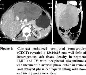
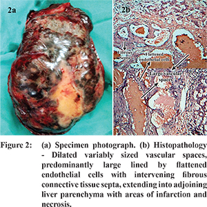
middle aged married female presented with complaints of mass per abdomen since 3 months and pain in the upper abdomen since 1 month. She had no history of jaundice or oral contraceptive use. On examination, a single swelling measuring 15x10 cm was found in the left hypochondrium and epigastrium crossing the mid line medially, up to the umbilicus inferiorly. Contrast enhanced computed tomography (CECT) revealed a 13x10x15 cms well defined heterogenous soft tissue density in segment II,III and IV with peripheral discontinuous enhancement in arterial phase, while in venous and delayed phase centripetal filling with non-enhancing areas were seen (Figure 1). The impression obtained on the CECT scan was of an atypical hemangioma. All the routine blood investigations were within normal limits except for anaemia. She was planned for left hepatectomy. Her abdomen was opened through a Mercedes benz incision. Introperatively, we found a 15x10x10 cms mass in the left lobe to the left of the falciform ligament. Compensatory hypertrophy of the right and caudate lobes was also observed. There was no evidence of portal hypertension. GIA 45 mm stapler was used for parenchymatous transection and left hepatic vein was ligated with prolene 3-0 suture. Histopathological examination revealed a cavernous hemangioma (Figure 2,2a).


